


The Neck
Description
This section is from the book "Lessons on Massage", by Margaret D. Palmer. Also available from Amazon: The Book Of Massage: The Complete Stepbystep Guide To Eastern And Western Technique.
The Neck
The Neck.—The vertebrae of the neck are seven in number. The two upper articular processes of the first vertebra are concave, and articulate with the occipital condyles, which are at each side of the foramen magnum, and which are convex. The occipital bone around the foramen magnum is attached by special ligaments to the anterior, posterior and lateral portions of the axis. The atlas is also attached to the occiput and to the axis by strong ligaments. Some of the ligaments are inside the spinal canal. The central odontoid ligament is attached to the tip of the odontoid peg, and to the anterior margin of the foramen magnum. The lateral odontoid, or check ligaments, are attached to each side of the peg, and to the inner surface of the occipital condyles. The transverse ligament behind the peg is attached to the sides of the atlas. The head is capable of forward flexion, extension, lateral flexion and rotation. The spinous processes of the cervical vertebrae are bifurcated, except the first and the last. The transverse processes are bifurcated, and each has a hole through which the vertebral artery passes, and is thus protected by a bony wall. The ligaments of the five lower cervical vertebrae are a continuation of the anterior and posterior common ligaments of the spine. The neural arches are bound together by ligamentâ subflava, interspinous and intertransverse ligaments.
The ligamentum nuchae, which reaches from the seventh cervical vertebra to the crest of the occipital bone, is a continuation of the supraspinous ligament ; it is attached by slips to all of the cervical vertebrae, its posterior border is free, and gives origin to the trapezius, rhomboid, serratus posticus superior, and splenius muscles.
The hyoid bone is shaped something like a horseshoe. It is under the lower jaw, between the root of the tongue and the larynx ; it has a base, and two hornlike processes at each side ; the longer horns or cornua go along the sides of the throat, the smaller are directed upwards, and are attached by ligaments to the styloid processes of the temporal bone. They keep the openings of the trachea and œsophagus extended. The chief muscles of the tongue and the trachea arise from the body of the bone ; the chief muscles of the oesophagus arise from the horns.
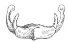
Fig. 91.—Hyoid Bone.
The neck contains the pharynx ; upper end of oesophagus ; larynx ; upper part of trachea ; external, internal, and anterior jugular veins ; external, internal, and common carotid arteries ; vertebral artery ; cervical plexus ; brachial plexus ; ganglia of sympathetic system ; phrenic, pneumogastric, spinal accessory, and hypoglossal nerve; apices of lungs; upper end of thoracic duct ; right lymphatic duct ; lymphatic glands ; numerous muscles.
The pharynx is at the back ; it extends from the skull to the fifth cervical vertebra ; the oesophagus is continuous with the pharynx. The larynx is in the middle of the neck, in front of the pharynx ; the trachea extends downwards from the larynx. Air to and from the larynx, as well as food from the mouth to the oesophagus, passes through the pharynx. The larynx is the organ of voice ; it consists of nine cartilages, held together by ligaments, and moved by muscles. The four largest cartilages, by which the vocal cords are supported, are the thyroid, cricoid, and arytenoids.
The thyroid is in the front ; it is funnel-shaped, and forms the prominence called Adam's apple.
The cricoid forms the lower part of the cavity, and is in shape like a signet-ring.
The two arytenoids are on each side at the back, on the upper border of the cricoid.
The epiglottis is a heart-shaped fibro-cartilage behind the root of the tongue, and in front of the superior opening of the larynx. In the act of swallowing this opening is closed by the folding forward of the two arytenoid cartilages, so that their apices are applied to the epiglottis, and thus solid or liquid matter is prevented from going into the trachea.
Continue to:


My Books

